Digital Anatomy Dissection Table – Advance Pro
IKAD: Transforming Education & Research with Powerful Equipment & Solutions. > Digital Anatomy Dissection Table – Advance Pro
Digital Anatomy Dissection Table - Advance Pro
Click to view full size image

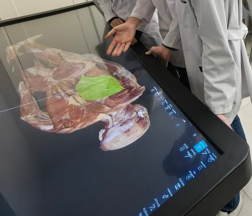
The II 88 Digital Anatomy Table represents a cutting-edge educational tool that leverages advanced digital 3D construction technology to create highly realistic, interactive, and detailed virtual representations of the human body. This system offers several key features that make it an innovative and powerful resource for teaching human anatomy. Below is a breakdown of its components and features:
Software Content
HUMAN GROSS ANATOMY
The 3D structure of the human body in the human anatomy module is digitally restored from male data (17000+ total layers of cross-section; resolution 13700*6340) and female data (16000+ total layers of cross-section; resolution 12000*5700) able to perform virtual anatomy operations through it.
HUMAN ANATOMY SYSTEM
The system contains a wealth of learning resources: more than 3000 3D structures, more than 3000 sectional images, more than 1700 images of CT/MRI, more than 100 teaching videos and more than 1800 exercises. Through the Anatomy Teaching System, teachers can operate professional anatomy teaching soft-ware through the touch pad. Students can watch stereo human anatomy structures by wearing 3D glasses, which can be viewed in an open, autonomous and interactive virtual environment Conduct an efficient, safe and economical teaching process.
EMBRYOLOGY
The system is a collection of videos, animations, 3D models and courseware as an integrated teaching platform.
The system is categorized in three modules on early human embryogenesis (General), the genesis of human embryonic organ systems (Monographs) and congenital malformations, each of which has the following teaching resources: preface, general video, knowledge points and exercise bank.
SLICE LIBRARY
The section library module contains at least 395 digital sections of histology and 780 digital sections of pathology. The slice library supports touch or mouse to simulate the operation under the mirror. One-click 4X, 10X, 20X, 40X objective lens magnification adjustment, also can pan to adjust the observation position, one click to select the history of browsing sections or favorite sections.
CLINICAL CASES
The clinical case module contains the number of real clinical cases not less than 180. It can display the disease name, basic information, chief complaint, image performance and diagnosis of the current case. The system provides window adjustment for CT/MRI images, and the window width and window position can be manually adjusted according to different parts so that users can quickly view different image contents.
Click to view full size image


Technology Features
- UHD HIGH-PRECISION REALISTIC ANATOMICAL STRUCTURE
- HIGH-PERFORMANCE ANATOMY TEACHING TOOL
- USER INTERACTION MANAGEMENT SYSTEM
- INTERACTIVE TOUCH CONTROL OPERATION
Hardware
-
Single Screen (Without Splicing): Size: 88-inches,
Resolution: 3840 * 1080; Brightness: 700cd/m2
Contrast: 1100:1
Touch mode: Infrared Touch. -
Computer:
I7 above generation 10 /64G DDR4 3200 / 4 T NVME SSD /RTX3080 above 10G /win10 -
Screen Electric Lifting:
The 88-inch display can be raised and lowered vertically and can be reversed 90 degrees.
Virtual 3D Construction of the Human Body
Realistic Anatomical Representation: The Digital Anatomy Table uses high-definition, high-precision 3D modeling to generate accurate, lifelike representations of the human body. These models are built from a variety of medical visualization resources, such as CT scans, MRI data, and other imaging techniques, allowing for a realistic and comprehensive anatomical structure.
Detailed Anatomy: The system includes detailed models of macroscopic (visible to the naked eye) and microscopic (cellular and tissue-level) anatomical structures. This allows users to explore the human body at various scales of detail.
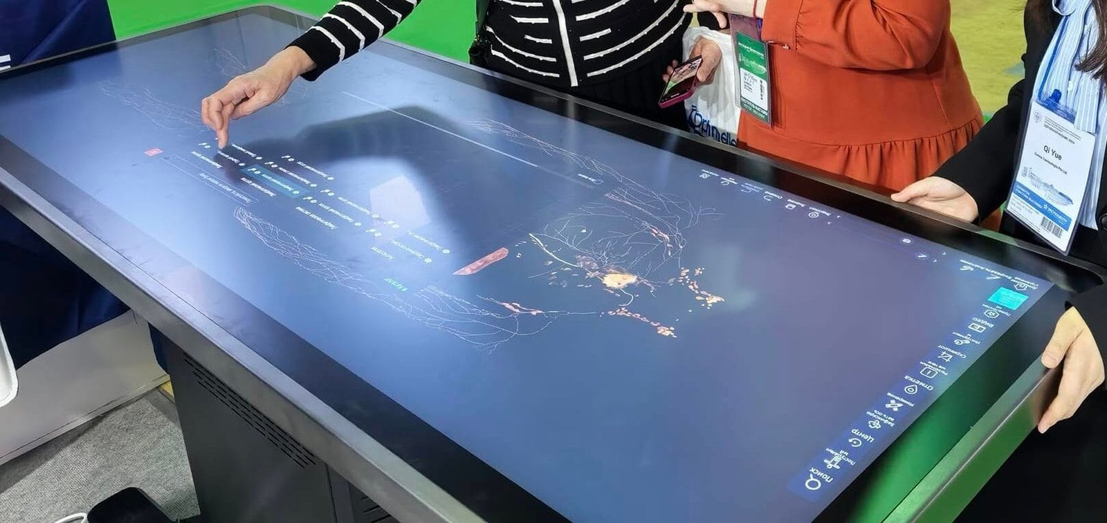
Interactive Touch Control Operation
User-Friendly Interface: The Digital Anatomy Table is equipped with a touchscreen interface that allows users to interact with the virtual body. Through touch control, users can:
Cut and Slice: The system allows users to “cut” or slice through the 3D model of the body in multiple directions and angles, enabling a closer inspection of internal structures.
Rotate and Zoom: Users can rotate the body to view structures from different angles or zoom in for a more detailed examination of specific organs, tissues, or systems.
Multi-Layer Exploration: The ability to observe the body at different layers (e.g., skeletal, muscular, vascular, etc.) provides a deeper understanding of how anatomical structures are organized and interact.
Integration of Anatomical and Sectional Knowledge
Comprehensive Learning: The system seamlessly integrates the knowledge of human anatomy (macroscopic structure) and sectional anatomy (cross-sectional images). This allows users to understand both the overall structure of the body and how it appears in slices or cross-sections, aiding in the understanding of real-world imaging techniques. Normal and Pathological Anatomy: The system provides the opportunity to study both healthy anatomy as well as diseased or abnormal anatomy. This makes it a valuable tool not only for general anatomical education but also for exploring specific conditions or pathologies.
System Integration and Visualization Resources
High-Performance Visualization: The system integrates high-performance rendering technologies to produce ultra-high-definition (UHD) anatomical models. This results in incredibly detailed and realistic visualizations, which are critical for accurate education and understanding.
Comprehensive Anatomical Resources & User Interaction Management: The system is designed to manage user interactions efficiently, allowing multiple users to explore the anatomy simultaneously or access specific content tailored to their level of expertise or learning needs.
Virtual Training Platform
Immersive Learning: The Digital Anatomy Table provides an immersive learning experience, combining high-quality digital anatomy with interactive features. This makes it an effective virtual training platform for students, medical professionals, and researchers, allowing them to explore complex anatomical structures and practice their skills without the limitations of physical cadavers or models.
3D Structure Exploration: Users can study the human body’s structure from various perspectives, including frontal, sagittal, and transverse views, enhancing their spatial understanding of anatomy.
Educational Benefits
Interactive and Engaging: The Digital Anatomy Table provides an engaging, interactive, and hands-on learning experience that encourages exploration and active learning. It is particularly useful in anatomy education, as students can manipulate the 3D models in real-time, enhancing their understanding of human body systems.Support for Multiple Learning Styles: The system caters to various learning styles—visual, tactile, and kinesthetic—by offering dynamic interaction with the 3D models, making it an effective tool for diverse groups of learners, including students and medical practitioners.
Advanced Teaching Tool: Teachers and professors can use the system to guide students through specific structures, clinical scenarios, or anatomical pathways, fostering a deeper understanding of complex concepts.
Application in Various Fields
Medical Education: The system is ideal for medical schools, universities, and teaching hospitals, where it can be used for teaching anatomy, pre-surgical planning, and clinical visualization.
Surgical Training: Surgeons can use the system for simulation and practice, gaining insights into anatomy relevant to their specialties, such as orthopedics, neurosurgery, or cardiology.
The Digital Anatomy Table offers a high-performance, interactive educational tool with advanced visualization and human-computer interaction technologies. It enhances the learning experience by allowing users to explore the human body at various scales, directions, and layers. Its ability to simulate real anatomical structures, and its user-friendly interface make it an invaluable resource for students, medical professionals, and educators alike, providing an immersive and flexible platform for anatomy learning and medical training.