Digital Anatomy Dissection Table – Advance
Digital Anatomy Dissection Table - Advance

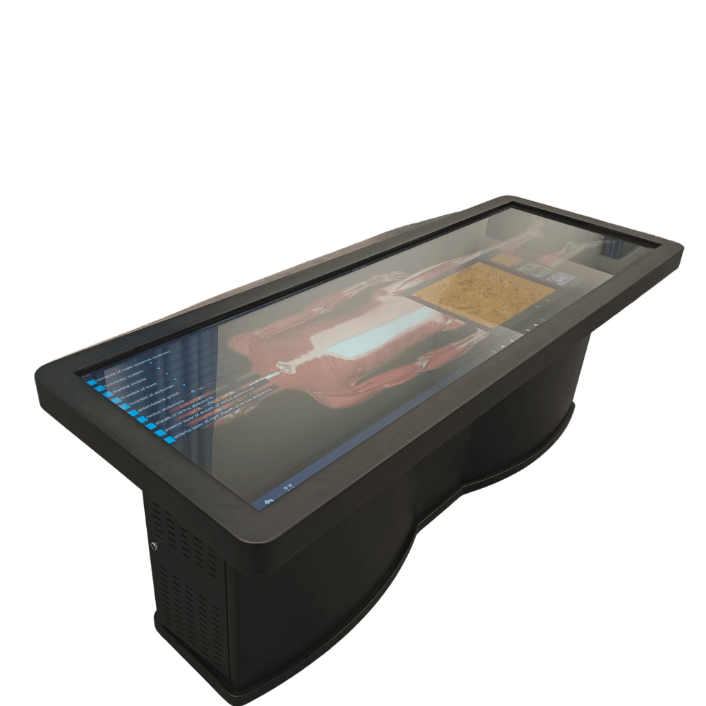
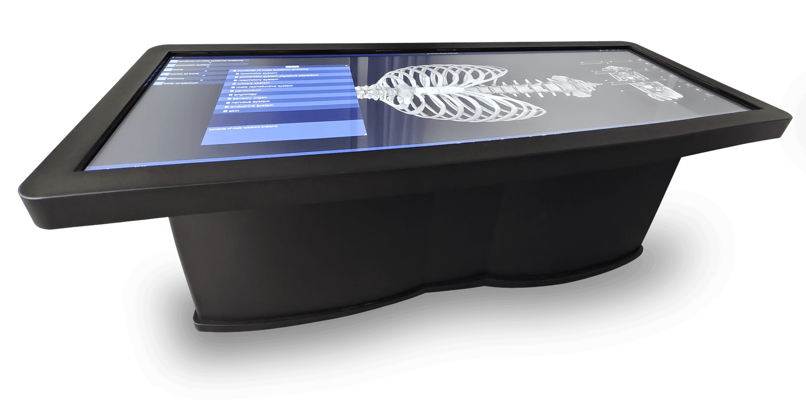
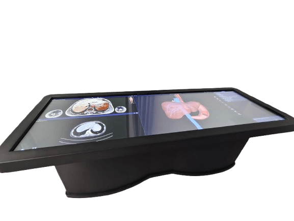
The Digital Anatomy Table categorizes anatomy into systematic anatomy, regional anatomy, and sectional anatomy, contains 6000+ 3D anatomical structures, bringing more detailed learning. Contains real dissection videos and anatomy animations to visualize the anatomy process. Provides a large number of test question resources to enhance knowledge consolidation.
The Digital Anatomy Table contains 2 sets of complete human tomographic image data for male and female: 2110 layers for male, precision 0.1mm-1mm; 3640 layers for female, precision 0.1mm-0.5mm, which can display more than 6000 anatomical structures in 3D form, and is the only digital anatomy teaching product based on the reconstruction of complete tomographic data.
The system covers systematic anatomy, regional anatomy, tomographic images, clinical cases, etc. It is equipped with corresponding CT and MRI images (more than 1700) on the basis of tomographic specimen images, and has more than 130 anatomy teaching micro-class videos and a large number of digital practice questions.
-
Screen: Single Screen (No Splicing): Size: 80-inches;
Resolution: 3840 * 1220;
Brightness: 700cd /m2;
Contrast: 1300:1;
Touch mode: infrared touch. - Computer: Processor I5; Graphics card 4G; Memory 8G; Hard disk 500G
- It has at least 3 USB ports and 1 HDMI and 1 VGA for displaying output on the screen.
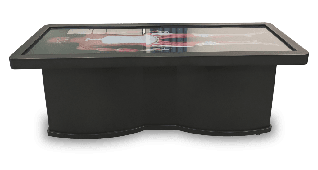
- The display screen is not less than 2260 x 707 x 750 millimeters (length x width x height), visual angle 89/89/89/89, weight approximately 185 kg.
- Dims : 2035x805.2x448.4mm
- (The hardware configuration will be adjusted accordingly with the product upgrade)
Click to view full size image